Swissy Health
We encourage educating yourself about Greater Swiss Mountain Dog health issues.
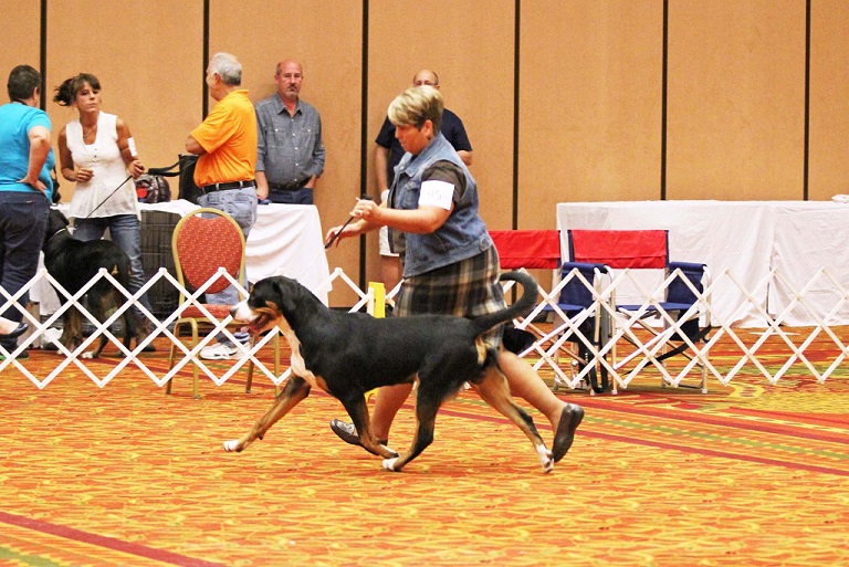
Photo Credit: Debbie Fields
A Note About Swissy Health
When choosing a Swissy breeder, you should aim to make a connection with a breeder who is completely transparent about any health issues AND their potential to appear in their breedings. Breeders who are seeking to further the breed as a whole will be meticulously mindful about the decisions to use dogs with known major health issues or a history of producing them. Swissys in general are prone to some disorders and issues, due to size, or widespread genetic history. Be very wary of a breeder who promises that issues have NEVER appeared in their breedings or in their dog's heritage.
Many issues, such as epilepsy, appear at some point in nearly all Swissy pedigrees. The ideal case is to find a breeder who is mindful about limiting the risk of major health issues, has thorough health histories of all dogs produced, and who is most of all honest and easily forthcoming about major health issues they have produced. If you are talking to a breeder who sidesteps around health issues or especially who denies any chance for epilespy in a breeding, I highly suggest looking elsewhere. This is simply dishonest. Please continue reading below to learn about breed-specific health issues.
Bloat (GDV)
Gastric Dilatation (GDV) is a very quickly progressing and life-threatening condition in dogs. This condition is generally linked to eating large meals, while many other possible causations are rumored. Whatever the cause, in occurences of GDV, the stomach dilates (because of food and/or gas expanding the stomach), and can escalate until neither food nor gas may be expelled from the stomach. The stomach continues to expand, and the pressure increases, as the causes of dilation cannot escape. That increased pressure and size of the stomach has several consequences, including:
- Prevention of adequate blood return to the heart from the abdomen;
- Loss of blood flow to the lining of the stomach;
- Rupture of the stomach wall;
- Pressure on the diaphragm preventing the lungs from adequately expanding, leading to decreased ability to maintain normal breathing.
The entire body suffers from this poor ventilation, leading to rapid cell death in many tissues. If the stomach continues dilating, there is potential for the stomach to rotate within the abdomen. This rotation can lead to blockage in the blood supply to the spleen and stomach. Most pets in this stage are in medical shock due to the effects systemwide.
The treatment for this condition includes stabilizing your dog, decompressing the stomach, and surgery to return the stomach to the normal position permanently. This surgery is known as gastropexy, in which the stomach is tacked to the abdominal wall to prevent future stomach rotation. Some Swissy owners choose to do this "stomach tacking" if/when the dog is already under anesthesia, such as during a spay/neuter, or any other procedure as a preventative measure. While mostly effective, the stomach can still rotate in the future, in some cases. Gastropexy lowers the risk of the rotation, but is not 100% preventative.
Osteochondrosis of the Shoulder in the Dog | Dog OCD | Joint Mouse. (2015, March 24). Retrieved October 9, 2015.
Splenic Torsion
Splenic torsion occurs when the spleen rotates or twists in the abdomen, preventing blood drainage and causing enlargement of the spleen. This condition is especially related to giant breed dogs with deep chests (such as Swissys). Splenic torsion can occur on its own or in combination with GDV.
Splenic torsion can be an acute condition manifested with pain or collapse and can be a life-threatening condition, or can be chronic associated with non-specific signs such as:
- Intermittent abdominal pain;
- Vomiting;
- Inappetence;
- Abdominal distention;
- Weight loss;
- Excessive drinking & urination;
Dogs are stabilized prior to surgery with fluids and blood, if necessary. Then, the spleen is surgically removed.
Osteochondrosis of the Shoulder in the Dog | Dog OCD | Joint Mouse. (2015, March 24). Retrieved October 9, 2015.
Epilepsy
First published in the January 2004 issue of the AKC Gazette & reproduced with permission from the author.
Epilepsy & the Greater Swiss Mountain Dog
Several serious heritable diseases may affect the Greater Swiss Mountain Dog and pose significant risk to the future health of our breed. Those of greatest concern are epilepsy, bloat, splenic torsion, and hip dysplasia. There are many factors related to epilepsy that make it a disease of great concern for Swissy fanciers.
The median age of death from bloat and splenic torsion in the GSMD is 9 years of age, largely due to available surgical options and treatment. However, the median age of death from epilepsy in the Swissy is 3.75 years. Though canine idiopathic epilepsy commonly develops after two years of age, the onset in Swissys may be much later. A late onset disease is of special concern to breeders, as a dog or bitch may be bred several times before being identified as a carrier or possibly even an affected animal. Unfortunately, there are no tests to determine if a Swissy carries these genes until it develops the disease, produces affected offspring, or until a parent develops the disease.
From an owner's perspective, epilepsy is an extremely challenging disease to manage. Oftentimes, Swissys do not respond well to drug therapy and continue to experience seizures and fatalities despite increasing medications. These medications have a high degree of toxicity and side effects, and require dogs to be carefully and continuously monitored. Management of an epileptic dog often severely impacts the lifestyle of the owner.
Canine genetics researcher, Dr. George Padgett, recently analyzed our health survey data and concluded that at least 39% of GSMDs carry the genes to produce epilepsy. A high priority must be placed on reducing this carrier rate in our breed. Sadly, even the most concerned breeder can produce puppies that develop epilepsy. Those who reveal cases of the disease and strive to eliminate carriers demonstrate a very high degree of ethical breeding.
Many matings carry an inherent risk to produce epilepsy. Those risks can be reduced if breeders utilize information about epilepsy in the relatives of prospective mates, and remove all known carriers from the breeding population. Offspring of affected dogs should not be bred, and care should be taken in selecting mates at low risk for carrying the genes for the disease. Healthy dogs known to have affected relatives should only be bred to dogs with good depth of normalcy in the pedigree. Making education on epilepsy a high priority is another key element in our effort to reduce this disease. Maintaining contact with homes and reporting cases of epilepsy to owners of littermates could positively affect future breeding decisions.
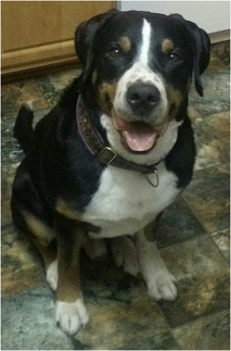
In loving memory of my special boy, Grainger.
Orthopedic & Joint Health
Prevention
As a giant breed dog, Swissys are more prone to joint and bone issues. It is recommended that you supplment your Swissy's diet with Glucosamine and Chondroitin to aid in the lubrication and strength of joints and bones. I recommend starting my puppies on GlycloFlex III around 18-24 months old and staying on it throughout their lifetimes. While many foods formulated for large dogs include these supplements, almost none of them contain actual therapeutic doses recommended for giant breed dogs. To help prevent major bone and joint damage, it’s important to avoid repetitive exercise on hard surfaces (running, biking, treadmills, jumping down, etc.) with your dog until the dog is at least 18-24 months old. Talk with your breeder about your specific dog before deciding at what age certain activities (and which) are safe for your Swissy. It’s very important to keep your dog at an appropriate weight to avoid excessive pressure on the joints, especially in a growing puppy or in an older adult.
The biggest way to prevent possible orthopedic concerns is to make sure the puppy’s parents and grandparents have their health clearances for hips, elbows, and shoulders. Genetic factors play a huge part. It is possible, however, for a dog to develop orthopedic problems even when the parents and grandparents are free of these problems because the current testing is not genetic – it’s phenotype testing only (meaning the tests only review the actual joints via xray – not the genetics of the dogs). Between good familial history for joint health, proper exercise and nutrition, and being careful with a growing puppy, you can set yourself up for the best possible outcome for your puppy’s joints as he matures.
Hip Dysplasia
Hip dysplasia refers to the development of a poorly-aligned joining of the femoral head and the acetabulum, allowing loose movement and varied pressure. These changes over time cause pain, joint damage, and inflammation. Fluid in the joint increases, and the round ligament that binds the femoral head to the acetabulum becomes enlarged. The normally smooth articular cartilage covering the end of the opposing femoral head and acetabulum is abraded and weakened, and the joint capsule becomes inflamed and thickened. Over time, the whole joint is structurally weakened and more painful.
Hip dysplasia is not easily observable in very young dogs. Joints of newborn puppies (who will later develop hip dysplasia), seem to be structurally sound.
Researchers have overcome one major hurdle by developing a test for hip dysplasia that is more accurate at younger ages than traditional procedures. Taking advantage of the wealth of information available from early detection, the next step is to gather and study DNA sequences in an effort to locate specific sequences that are markers for either normal or abnormal hip joint development. This challenging task is made more difficult by the fact that there are almost certainly a number of different genes that trigger hip dysplasia, but the research team is confident that, by utilizing ever-increasing molecular technologies, they will soon crack the genetic code of hip dysplasia. Dr. Rory Todhunter, Professor of Surgery at the Cornell University College of Veterinary Medicine, and his team of researchers have successfully used an experimental breeding program of Labradors (high risk for hip dysplasia) and Greyhounds (low risk) to identify chromosomal regions harboring genes that confer susceptibility to and protection against hip dysplasia.
Further, though the DLS test is significant improvement upon traditional diagnostic measures, there is a continuing search for the metabolic causes of hip dysplasia that work in conjunction with genetics. It is noteworthy that despite its genetic basis, the development of hip dysplasia is also influenced by environments. Topics such as the possible metabolic abnormalities are areas that Institute researchers continue to explore.
Baker Institute | Animal Health. (n.d.). Retrieved October 9, 2015.
Elow Dysplasia
Canine elbow dysplasia (ED) is a disease of the elbows of dogs caused by growth disturbances in the elbow joint. There are a number of theories as to the exact cause of the disease that include defects in cartilage growth, trauma, genetics, exercise, diet and so on. It is likely that a combination of these factors leads to a mismatch of growth between the two bones in the fore leg located between the elbow and the wrist (radius and ulna). If the radius grows more slowly than the ulna it becomes shorter leading to increased pressure on the medial coronoid process of the ulna (Figure 1). This in turn can cause damage to the cartilage in joint and even fracture of the tip of the coronoid process, which damages the medial compartment (side closest to the body) of the joint. Less commonly, if the ulna grows too slowly then the radius pushes the upper arm bone (humerus) against the anconeal process, which can then lead to failure of the anconeal process to attach to the ulna at maturity. It is believed that the mismatch in growth between the radius and ulna may sometimes only occur during a puppy’s growth, but it may also persist when the pup has finished growing.
Elbow dysplasia is most often seen in large to giant breed dogs, particularly Labradors, Golden Retrievers, German Shepherds, and Rottweilers, but can occur in most breeds of dog. Different breeds have predispositions to different forms of the disease, so that ununited anconeal process (UAP) is largely a problem of German Shepherds, medial compartment disease (medial coronoid injury) is seen in many other breeds and sight hounds are free of the disease.
Unfortunately once the elbow joint has been damaged through either cartilage loss, the presence of medial coronoid fragments or an ununited anconeal process, a vicious circle of inflammation and further cartilage damage begins. Ultimately this causes progressive arthritis of the elbow joint leading to pain and loss of function.
Dogs affected by elbow dysplasia often show signs from an early age, typically from 5 months on, but some may first be diagnosed after 4–6 years. Affected dogs develop a front limb lameness that typically worsens over a period of weeks to months. Lameness is usually worse after exercise and typically never completely resolves with rest. Often both fore legs are affected, which can make detection of lameness difficult, as the gait is not asymmetric. When both elbows are involved the dog usually becomes unwilling to exercise for long periods or may even refuse to complete a walk.
Treatment depends on the severity of the disease in the elbow. In many cases surgery is recommended, but your veterinarian may recommend medical management if the problem is very mild or so severe that the joint is not likely to benefit from routine surgery. Treatment can be divided into the correction of a joint step between the radius and ulna if present, and treatment of any other joint damage. Often surgery is best performed arthroscopically, but conventional open surgery can also be done. Depending on the individual dog’s elbow problem surgery may involve:
- Correction of joint step; is usually done by cutting the ulna to re-establish elbow congruence;
- Removal of any coronoid fragments and removal of loose cartilage
- Surgical alteration of the elbow joint to shift weight away from damaged areas
- Reattachment or removal of an united anconeal process of the medial joint compartment
- Joint replacement if the elbow is severely diseased
- Age;
- Gender;
- Breed (genetic);
- Rapid growth;
- Nutrient excesses (primarily calcium excesses);
Canine Elbow Dysplasia. (2015). Retrieved October 9, 2015.
Osteochondrosis
Osteochondrosis occurs commonly in the shoulders of immature, large, and giant-breed dogs. The lesion appears on the caudal (back) surface of the humeral head. Osteochondrosis begins with a failure of cartilage to form bone in the humeral head. This failure leads to abnormal cartilage thickening. Increased cartilage thickness may result in malnourished cartilage cells that die. Loss of these cartilage cells deep in the cartilage layers leads to formation of a defect at the junction between cartilage and bone. Subsequently, normal activity may cause fissures in the cartilage that eventually communicate with the joint, forming a cartilage flap. It is upon this flap formation that osteochondrosis becomes osteochondritis dissecans (OCD). OCD is the form of osteochondrosis that is associated with pain and dysfunction. In some cases, the resulting flap occupies as much as half the humeral head. The cartilage flap may completely detach from the underlying bone and become lodged in the back of the joint pouch. Free cartilage flaps can lodge in joints and may increase in size with calcification becoming joint “mice” which can be seen on radiographs.
The causes of OCD are multifactorial with genetic and nutritional interactions implicated. Risk factors for OCD include:
Due to the high frequency of occurrence within certain breeds of dogs and within certain bloodlines, hereditary may be a factor. Males are more commonly affected than females.
Clinical signs often develop when the dog is between four and eight months of age. Dogs usually begin limping on one of their forelimbs. In many cases, a gradual onset of lameness improves after rest and worsens after exercise. Although your pet may be lame on only one leg, this condition may be present in the opposite leg too.
Diagnosis of OCD is based on seeing a defect (flattening) present in the humeral head on X-rays of the shoulder. Radiograph both shoulders because this condition may be present in both shoulders, despite apparent lameness in only one limb. Often, older dogs that have had the problem for a while have large calcified joint mice.Surgical treatment involves removal of the cartilage flap from the joint and scraping the edges of the bony defect to ensure removal of all affected cartilage. Arthroscopy, a minimally invasive procedure using a very small tube with an attached camera best accomplishes this. Dogs with persistent lameness that is unresponsive to medical treatment may require surgical treatment.
Osteochondrosis of the Shoulder in the Dog | Dog OCD | Joint Mouse. (2015, March 24). Retrieved October 9, 2015.
Bleeding Disorders
While there is not much data available about this particular genetic mutation, there is a test for the P2Y12 mutation (discussed below) available. I’m not convinced the mutation ALONE results in a dog having otherwise unexplained excessive bleeding; however, until further information is available, I will continue to test my dogs and make reasonable breeding decisions using those test results. Both active bitches in my breeding program are P2Y12 CLEAR (they do not have the mutation).
A platelet disorder has recently been identified in Greater Swiss Mountain Dogs at the functional and molecular level. The first dog documented to have the disorder bled excessively following a routine spay.
Platelets are small, circulating cytoplasmic fragments that are the first line of defense in stopping the flow of blood from injured blood vessels. An important aspect of platelet function is their ability to stick to each other and plug holes in damaged vessels until blood clotting and tissue repair can occur. The platelets in affected Greater Swiss Mountain dogs are unable to respond properly to a specific platelet activating agent because of a dysfunctional or missing receptor. Therefore, these dogs are at increased risk for spontaneous hemorrhage and they are also at high risk for excessive hemorrhage as a result of injury or surgery. Post operative hemorrhage may be life threatening.
The types of spontaneous bleeding that may occur include excessive gingival bleeding during tooth eruption, nose bleeds, and superficial skin bleeds. By using DNA testing, affected and carrier animals can be identified by submitting a blood sample through the mail. Carrier detection is vital in controlling spread of inherited defects and DNA testing is the only reliable method of detecting these animals.
P2Y12 Receptor Platelet Disorder in Greater Swiss Mountain Dogs. (n.d.). Retrieved October 9, 2015.
Urinary Incontinence
The second most common health problem experienced by this breed is urinary incontinence. Many Greater Swiss Mountain Dogs urinate involuntarily, especially while sleeping. Although males can be affected, this condition is much more frequent in females, and 17% of all females suffer from this condition to varying degrees. This condition is much more likely to appear in spayed females than unsprayed ones. Allowing a female to go through at least one reproductive cycle can reduce the likelihood of her developing urinary incontinence, but it is no guarantee.